Thursday, December 17, 2009
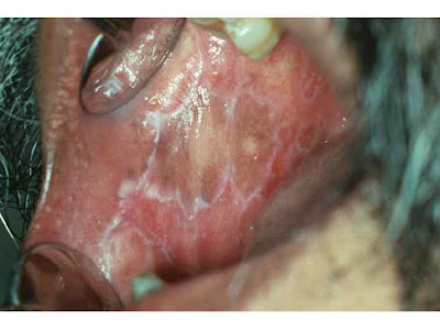
34 - Wickham's striae
Lichen planus in the right buccal mucosa. Note the lacy white Wickham striae and the localized hyperpigmentation.
*Wickham striae are whitish lines visible in the papules of lichen planus and other dermatoses, typically the macroscopic appearance of the histologic phenomenon hypergranulosis, and named for Louis Frédéric Wickham.
Thursday, October 22, 2009
33 - Kerion
A kerion is not an infectious agent in itself rather a kerion is the skin leison that develops when an infectious agent that normally causes scalp ringworm (tinea capitis) becomes more aggressive. Deep boggy red areas characterized by a severe acute inflammatory infiltrate with pustule formation are termed kerions or kerion celsi. Normally, scalp ringworm inducing agents cause circular patches of red crusty skin. Although not pleasant, the problem is relatively mild and reversible with proper treatment. However, if the infection gets out of hand a kerion may develop.
When a kerion develops it first starts out as a more typical presentation of tinea capitis with a flaky, crusty patch of skin on the scalp. This can quickly deteriorate into a boggy, puritic mass of inflamed tissue. This is a kerion. The kerion can deteriorate to a nasty, deep abscess if it is not treated correctly. This has the potential to to elicit scarring and permanent alopecia. When a kerion develops with severe inflammation it is fairly common for the regional lymph nodes in the neck to become enlarged (called cervical lymphadenopathy). Suppuration and kerion formation are more commonly are typically associated with Trichophyton tonsurans (dermatophytes) infection. Kerion formation with other infectious agents that cause tinea capitis are less likely, but still possible.
Sometimes Kerion Celsi is confused with other conditions due to a lack of diagnostic testing. In one report, some patients were hospitalized with the diagnosis of Staphylococcal abscess while a microbiological diagnostic test would have shown the cause was due to Trichophyton verrucosum infection resulting in a kerion (Zaror, 1995). Occasionally, a kerion can look much like some forms of scarring alopecia such as dissecting cellulitis or erosive pustular dermatosis. Because of this apparent similarity in presentation, the doctor needs to be very careful in their investigation and knowledgeable about the potential for confusing kerions with other diagnoses. The patient who suspects they have a kerion caused by an infectious agent or something similar needs to find an experienced dermatologist to improve the chances of getting a correct diagnosis. The average general practitioner is probably not in a position to make a kerion diagnosis with confidence.
Early initiation of treatment is extremely important with kerions. The sooner treatment is started the less likely the kerion will promote a permanent scarring alopecia. Unfortunately, some reports suggest that rapid diagnosis and treatment of kerions only occurs in a minority of cases or that kerions are often misdiagnosed. There seems to be a certain lack of knowledge about tinea capitis and kerions among some general practitioners and this can lead to a delay in receiving proper treatment. The longer kerions persist the more damaging they become. When a kerion is diagnosed, the typical immediate treatment response is a course of "Griseofulvin", an anti fungal agent. Most patients with kerions and a primary diagnosis of tinea capitis also have a secondary bacterial infection of the kerion. Griseofulvin is not good for treating bacterial or yeast infections so other anti-bacterial treatments may be given along with the Griseofulvin. Some published case reports have indicated the newer anti fungal agents Itraconazole and Terbinafine have also been successfully used to treat kerions. Sometimes oral corticosteroids are also given in addition to the anti fungal agent, although the few published studies comparing treatments with and without corticosteroids have shown little added benefit. However in principle, oral corticosteroids should help reduce the inflammation in the kerion. Topical corticosteroids are not used as this can complicate the local fungal infection.
Wednesday, April 22, 2009
32 - Cutaneous melanoma Risk factors
Monday, March 16, 2009
31 - AIIMS november 2001 dermatology mcqs with answers
1q: acne vulgaris is due to involvement of ?
a. sebaceous gland
b. pilosebaceous gland
c. eccrine gland
d. apocrine gland
2q: a patient with psoriasis was started on systemic steroids. After stopping treatment ,patient developed generalized pustules all over his body . what is the most likely cause ?
a. drug induced reactions
b. pustular psoriasis
c. bacterial infections
d. septicemia
3q: a patient presents with scarring alopecia, thinned nails, hypopigmentation, muscular lesions over trunk and oral mucosa. The diagnosis is ?
a. psoriasis
b. leprosy
c. lichen planus
d. pemphigus
4q: a young boy presented with lesion over his right buttock which had peripheral scaling and central scarring. The investigation of choice would be ?
a. tzanck smear
b. KOH preparation
c. Biopsy
d. Saboraud’s agar
30 - AIIMS may 2002 dermatology mcqs with answers
1q: treatment of pustular psoriasis is ?
a. thalidomide
b. retinoids
c. hydroxyurea
d. methotrexate
2q: a patient presents with erythematous scaly lesions on extensor aspect of elbows and knee. The clinical diagnosis is got by ?
a. auspitz sign
b. KOH smear
c. Tzanck smear
d. Skin biopsy
3q: actinic keratosis is seen in ?
a. basal cell carcinoma
b. squamous cell carcinoma
c. malignant melanoma
d. epithelial cell carcinoma
4q: a 30 year old female presents with history of itching under right breast . on examination annular ring lesion was present under the breast. The diagnosis is ?
a. trichophyton rubrum
b. candida albicans
c. epidermophyton
d. microsporum
5q: wood’s lamp light is used in the diagnosis of ?
a. tinea capitis
b. candida albicans
c. histoplasma
d. Cryptococcus
6q: a patient presented with multiple nodulocystic lesions on the face . the drug of choice is ?
a. retinoids
b. antibiotics
c. steroids
d. UV light
29 - AIIMS november 2002 dermatology mcqs
1q: a 45 year old man has multiple grouped vesicular lesions present on the T 10 segment dermatome associated with pain. The most likely diagnosis is ?
a. herpes zoster
b. dermatitis herpetiformis
c. herpes simplex
d. scabies
2q: a 28 year old patient has multiple grouped papulovesicular lesions on both elbows,knees,buttocks and upper back associated with severe itching. The most likely diagnosis is ?
a. pemphigus vulgaris
b. bullous pemphigoid
c. dermatitis herpetiformis
d. herpes zoster
3q: a child has multiple itchy popular lesions on the genitalia and fingers .similar lesions are also seen in younger brother. Which of the following is the most possible diagnosis ?
a. popular urticaria
b. scabies
c. atopic dermatitis
d. allergic contact dermatitis
4q: which layer of epidermis is underdeveloped in the very low birth weight infants in the initial 7 days ?
a. stratum germinativum
b. stratum granulosum
c. stratum lucidum
d. stratum corneum
5q: scabies an infection of the skin caused by sarcoptes scabie is an example of ?
a. water borne disease
b. water washed disease
c. water based disease
d. water related disease
Saturday, March 14, 2009
28 - common dermatological terms and their definitions
1. alopecia : hair loss , it may be partial or complete .
2. annular : ring shaped lesions
3. cyst : a soft, raised, encapsulated lesion filled with sesamoid or liquid contents .
4. herpetiform : grouped lesions
5. lichenoid : violaceous to purple, polygonal lesions that resemble those seen in lichen planus
6. milia : small ,firm, white papules filled with keratin
7. morbilliform : generalized , small erythematous macules and/or papules that resemble lesions seen in measles
8. nummular : coin shaped lesions
9. poikiloderma : skin that displays variegated pigmentation ,atrophy and telangiectases .
10. polycyclic : a configuration of skin lesions formed from coalescing rings or incomplete rings
11. pruritis : a sensation that elicits the desire to scratch. Pruritis is often the predominant symptom of inflammatory skin diseases ( example : atopic dermatitis , allergic contact dermatitis ) . it is also commonly associated with xerosis and aged skin . systemic conditions that can be associated with pruritis include chronic renal disease, cholestasis, pregnancy, malignancy, thyroid disease, polycythemia vera and delusions of parasitosis .
28 - secondary skin lesions description
1. lichenification : a distinctive thickening of the skin that is characterized by accentuated skin-fold markings
2. scale : excessive accumulation of stratum corneum
3. crust : dried exudate of body fluids that may be either yellow (that is serous crust) or red (that is hemorrhagic crust).
4. erosion : loss of epidermis without an associated loss of dermis
5. ulcer : loss of epidermis and atleast a portion of the underlying dermis
6. excoriation : linear, angular erosions that may be covered by crust and are caused by scratching .
7. atrophy : an acquired loss of substance . in the skin , this may appear as a depression with intact epidermis ( that is loss of dermal or subcutaneous tissue ) or as sites of shiny ,delicate ,wrinkled lesions ( that is epidermal atrophy )
8. scar : a change in the skin secondary to trauma or inflammation. Sites may be erythematous, hypopigmented or hyperpigmented depending on their age or character . sites on hair-bearing areas may be characterized by destruction of hair follicles .
Thursday, March 12, 2009
27 - primary skin lesions description
PAPULE : a small, solid lesion, less than 0.5 cms in diameter , raised above the surrounding skin surface and hence palpable . ( example : a closed comedone or whitehead, in acne ).
TUMOR : a solid, raised growth greater than 5 cms in diameter .
VESICLE : a small, fluid-filled lesion , less than 0.5 cm in diameter , raised above the plane of the surrounding skin. Fluid is often visible , and the lesions are translucent{example : vesicles in allergic contact dermatitis caused by toxicodendron (poison ivy)}.
BULLA : a fluid filled ,raised , often translucent lesion greater than 0.5 cm in diameter .
TELANGIECTASIA : a dilated , superficial blood vessel .

